The Battle Against Coronavirus (COVID-19). The Molecular Basis of Spirituality Science.
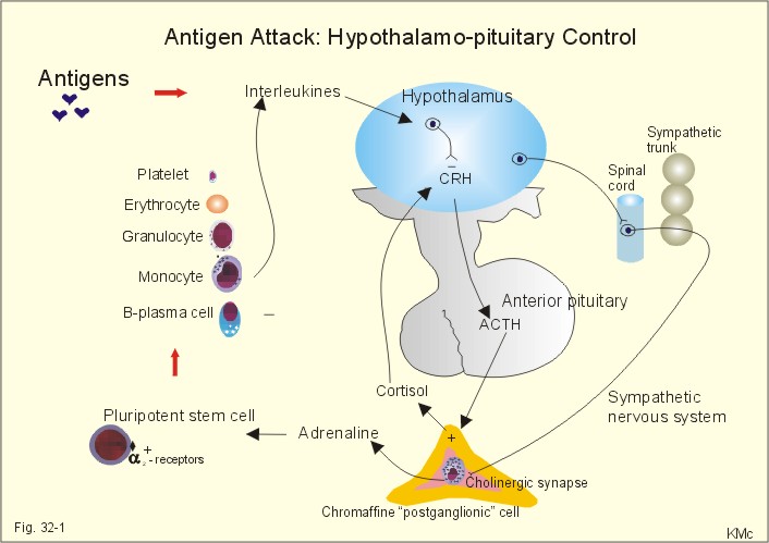
To account for human existence, to face challenges to human survival, man has to identify molecules that operate various living functions. Man defends his life deploying protein molecules that recognize and respond to invasion of human body by pathogens consisting of foreign protein molecules. While learned experience provides cortical awareness of human immunological responses to attacks by pathogens, human mind is blissfully unaware of presence of pathogens. Recognition of non-self proteins and molecules called antigens is not a mental function. If Spirit or Soul functions as guiding mechanism, Spirit or Soul will have awareness of invasion of body by foreign antigens and body’s immunological response to such invasion. If Reticular Formation of Brain Stem, the site at which contents of Consciousness are composed, is viewed as seat of human Spirit or Soul, it has awareness of body’s invasion and response to attacks by pathogens. Immunological responses to infections trigger a stress response mediated by Hypothalamus – Pituitary – Adrenal Cortex Pathway.
The discovery of antibiotic-resistant Superbugs and the discovery of protein structures of the Novel Coronavirus (COVID-19) may eventually lead to better understanding of Spirit or Soul in shaping human survival while warding off invasion by pathogens.
First 3D map of coronavirus protein paves way for vaccine design
This article, First 3D map of coronavirus protein paves way for vaccine design, originally appeared on CNET.com.
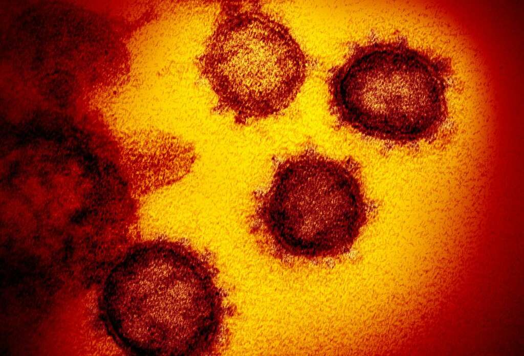
The first 3D map of SARS-CoV-2, the coronavirus responsible for over 2,000 deaths since December 2019, has been produced by a collaboration of coronavirus researchers at the University of Texas at Austin and the National Institutes of Health. Heralded as a breakthrough, the map provides a stepping stone to the development of antivirals or vaccines to stymie the virus.
In under two months, infections of the novel coronavirus have soared past 75,000 and caused significant economic turmoil in China and abroad. The World Health Organization has declared the outbreak a public health emergency of international concern and when it announced the name of the viral disease on Feb. 11, it added that a vaccine was likely 18 months away.
But scientists are mobilizing their resources quickly, sharing information about the virus with unprecedented pace, and turning experiments into peer-reviewed research in a matter of weeks.
That’s the case for Jason McLellan, a structural biochemist, and his team at UT Austin who have been studying similar coronaviruses for years. Their latest study, published in the journal Science on Wednesday, took advantage of state-of-the-art technology at the university to map the molecular structure of SARS-CoV-2, with a particular focus on the virus’ “spike protein.” The protein is critical to the viruses survival because it enables it to get inside human cells and begin making copies of itself. But what makes it dangerous also makes it a target.
The chief function of a vaccine is to prime the immune system. They work by presenting small parts of harmful pathogens like viruses and bacteria to our army of immune cells. It’s like a molecular “WANTED!” poster — the immune system gets a good look at any nasty bugs and starts to keep watch. If the real virus or bacteria sneak into the body, the immune system is ready and sends out an army of cells and antibodies to stop the invader.
After Chinese researchers shared the genetic sequence of the virus in January, the team were able to design and produce samples of the spike protein in the lab. Using a specialized form of microscopy, they then mapped its structure.
The research team showed there are similarities in the way the spike proteins work between the coronavirus responsible for the 2002-2003 SARS outbreak and the novel virus, SARS-CoV-2. However, the latter appears to bind to human cells much more strongly than the SARS virus did and antibodies against the first SARS virus don’t seem to react to the new virus in the same way.
Nevertheless, creating the 3D atomic scale map of the spike protein in SARS-CoV-2, the team were able to show it can elicit an immune response, making it a viable molecule to speed up vaccine design and development.
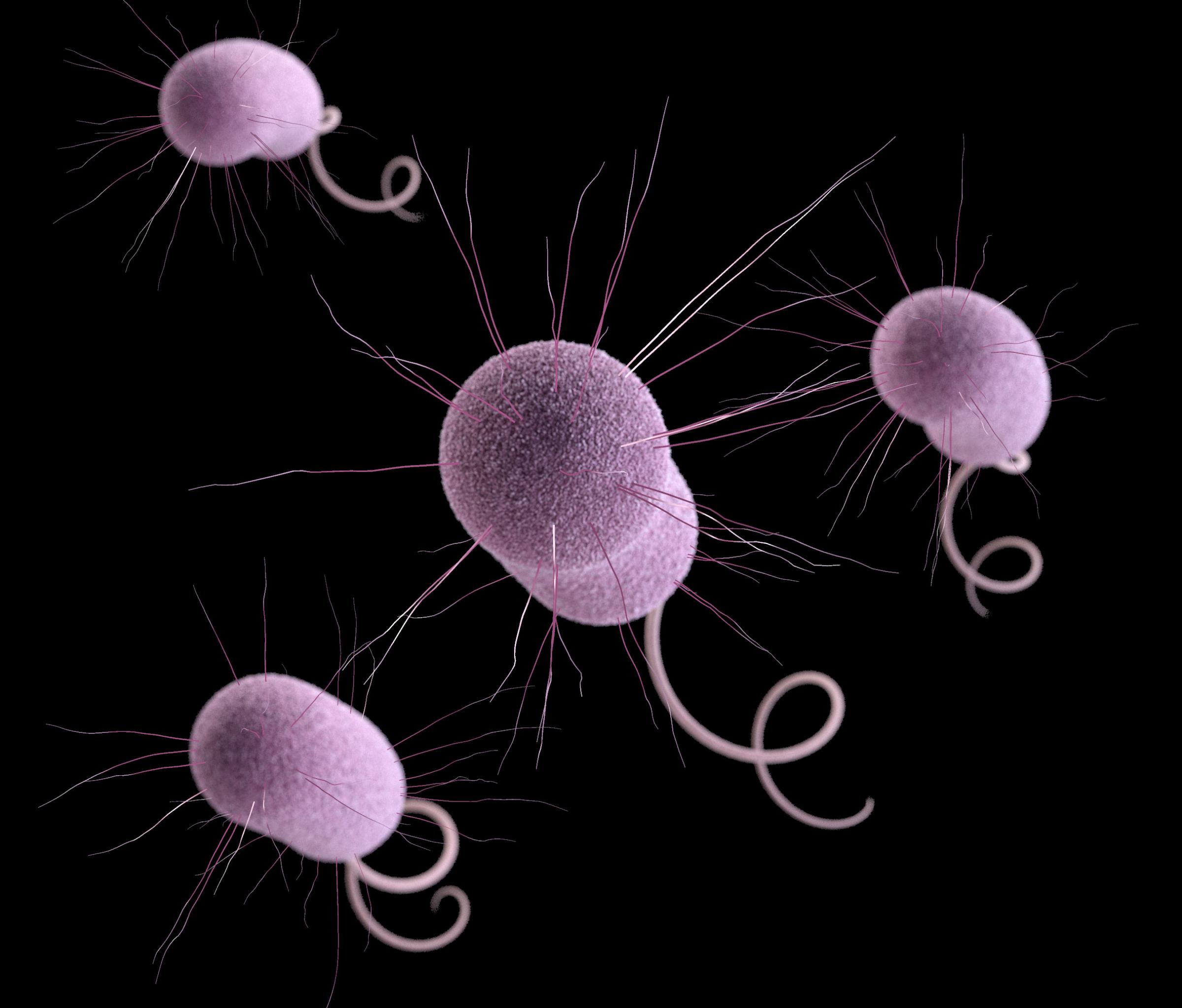
The coronavirus uses this protein to invade human cells.
Previous research revealed that coronaviruses invade cells through so-called “spike” proteins, but those proteins take on different shapes in different coronaviruses. Figuring out the shape of the spike protein in SARS-Cov-2 is the key to figuring out how to target the virus, said Jason McLellan, senior author of the study and an associate professor of molecular biosciences at the University of Texas at Austin.
Though the coronavirus uses many different proteins to replicate and invade cells, the spike protein is the major surface protein that it uses to bind to a receptor — another protein that acts like a doorway into a human cell. After the spike protein binds to the human cell receptor, the viral membrane fuses with the human cell membrane, allowing the genome of the virus to enter human cells and begin infection. So “if you can prevent attachment and fusion, you will prevent entry,” McLellan told Live Science. But to target this protein, you need to know what it looks like.
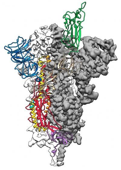
(Image: © Jason McLellan/Univ. of Texas at Austin)
Earlier this month, researchers published the genome of SARS-Cov-2. Using that genome, McLellan and his team, in collaboration with the National Institutes of Health (NIH), identified the specific genes that code for the spike protein. They then sent that gene information to a company that created the genes and sent them back. The group then injected those genes into mammalian cells in a lab dish and those cells produced the spike proteins.
Next, using a very detailed microscopy technique called cryogenic electron microscopy, the group created a 3D “map,” or “blueprint,” of the spike proteins. The blueprint revealed the structure of the molecule, mapping the location of each of its atoms in space.
“It’s impressive that these researchers were able to get the structure so quickly,” said Aubree Gordon, an associate professor of epidemiology at the University of Michigan who was not a part of the study. “It’s a very important step forward and may help in the development of a vaccine against SARS-COV-2.”
Stephen Morse, a professor at Columbia University’s Mailman School of Public Health who was also not a part of the study agrees. The spike protein “would be the likely choice for rapid development of vaccine antigens” and treatments, he told Live Science in an email. Knowing the structure would be “very helpful in developing vaccines and antibodies with good activity,” as would producing higher quantities of these proteins, he added.
The team is sending these atomic “coordinates” to dozens of research groups around the world who are working to develop vaccines and drugs to target SARS-CoV-2. Meanwhile, McLellan and his team hope to use the map of the spike protein as the basis for a vaccine.
When foreign invaders, such as bacteria or viruses, invade the body, immune cells fight back by producing proteins called antibodies. These antibodies bind to specific structures on the foreign invader, called the antigen. But producing antibodies can take time. Vaccines are dead or weakened antigens that train the immune system to create these antibodies before the body is exposed to the virus.
In theory, the spike protein itself “could be either the vaccine or variants of a vaccine,” McLellan said. When you inject this spike-protein-based vaccine, “humans would make antibodies against the spike, and then if they were ever exposed to the live virus,” the body would be prepared, he added. Based on previous research they did on other coronaviruses, the researchers introduced mutations, or changes to create a more stable molecule.
Indeed, “the molecule looks really good; it’s really well behaved; the structure kind of demonstrates that the molecule is stable in the correct confirmation that we were hoping for,” McLellan said. “So now we and others will use the molecule that we created as a basis for vaccine antigen.” Their colleagues at the NIH will now inject these spike proteins into animals to see how well the proteins trigger antibody production.
Still, McLellan thinks a vaccine is likely about 18 to 24 months away. That’s “still quite fast compared to normal vaccine development, which might take like 10 years,” he said.
The findings were published today (Feb. 19) in the journal Science.

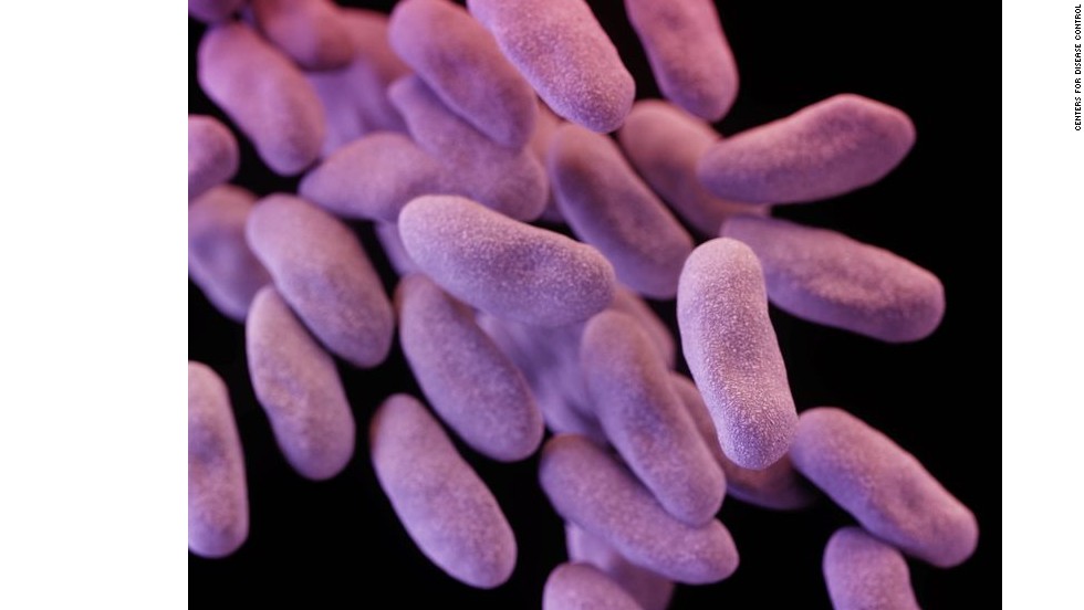
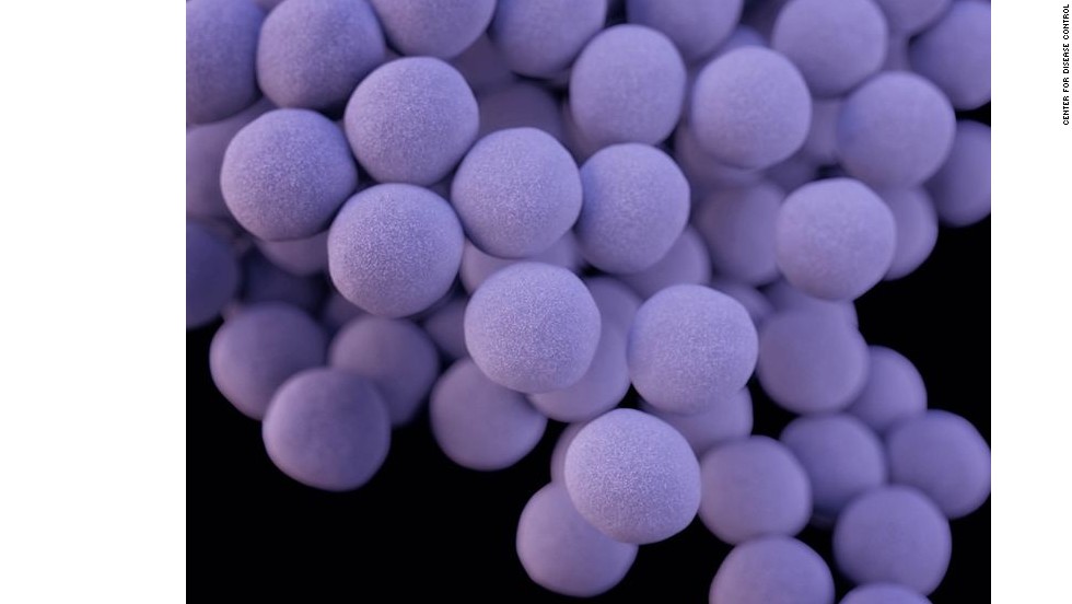
These are the top superbug threats in the U.S.

THE WHITE HOUSE HAS MADE THE GERMS ARE SERIOUS PRIORITY
Urgent threat. According to the White House plan to combat antibiotic-resistant bacteria, carbapenem-resistant Enterobacteriacea, CRE, is one of the country’s most urgent threats. Forty-four states have had at least one type of confirmed CREcase, which are resistant to nearly all antibiotics including last-resort drugs. CDC
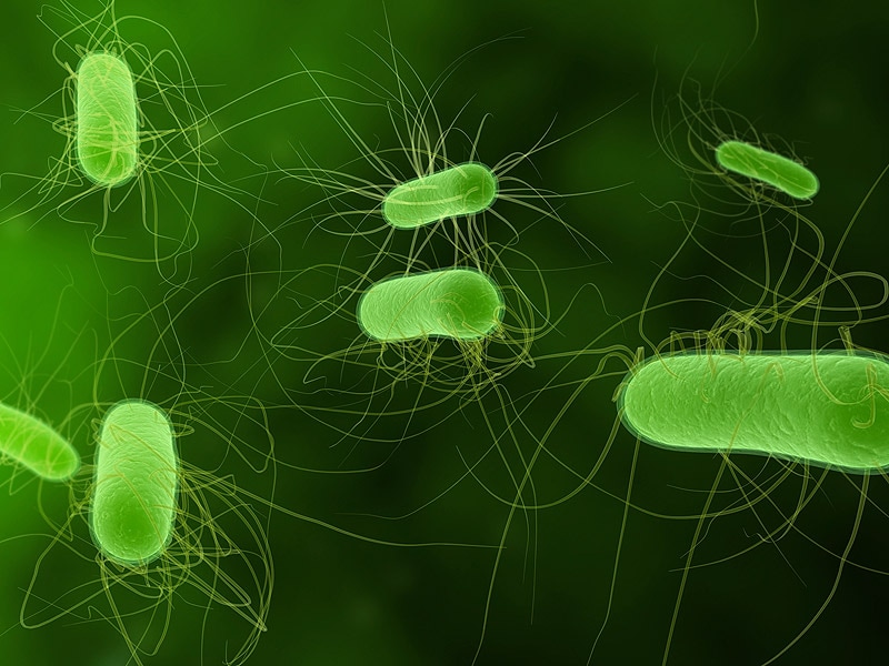
Meanwhile, another surveillance system that includes the CDC, the USDA and the Food and Drug Administration has also been searching for the gene in bacteria collected from food animals, meat sources and people. Scientists have scoured more than 44,000 samples of salmonella bacteria and 9,000 samples of E. coli and Shigella bacteria.
That search is how the USDA scientists recently found the gene in a sample from a pig intestine. It also was in a strain of E. coli, and also on a plasmid. The USDA is working to determine the sample’s origin.
The strains and plasmids appear to be different, McGann said. That suggests that the gene is circulating through at least two — and possibly more — routes within the United States.
McGann said he learned about the mcr-1 gene in the pig sample only when his team notified government officials about its own finding. U.S. officials haven’t provided details about when the animal sample was found or why information about it wasn’t disclosed earlier.
Public health officials are most worried about the colistin-resistant gene spreading to a family of superbugs known as CRE, for carbapenem-resistant enterobacteriaceae, which the CDC has called one of the country’s most urgent public health threats. In some instances, CRE kills up to 50 percent of patients who become infected. Colistin is increasingly the last-resort drug to treat patients with such infections.


No comments:
Post a Comment